CUNJC’s Project Collaboration Publishes in Nature Communications
Functional biological living material is a new type of composite material, which aims to attach functional microorganisms to traditional materials, so that the material has certain biological functions, such as biocatalysis, biodegradation, and biomanufacturing. How to precisely manipulate the formation of this material in space while controlling the distribution of cells and the microenvironment is a major problem in this field.

In response to this problem, Professor Yu Ziyi from Nanjing Tech University, also the PI of CUNJC’s research project "Engineering and Biological Application of Droplet Microfluidic Platform", and the research team of Professor Tuomas Knowles of the Department of Chemistry of the University of Cambridge, developed a new type of bioink. The ink is composed of hydrogel microspheres and utilizes the liquid-liquid phase separation behavior between two biomaterials to form a core-shell structure, separating the microenvironment of cell growth from the 3D printing of the material.
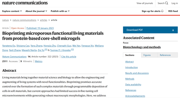
The research result, titled "Bioprinting microporous functional living materials from protein-based core-shell microgels", was published in the international high-level academic journal Nature Communications on 19 January. Ou Yangteng, a doctoral candidate in the Department of Chemistry of Cambridge University and a Research Assistant of CUNJC, is the first author. Professor Yu Ziyi and Professor Tuomas Knowles are the corresponding authors. This paper is one of the joint research achievements of the University of Cambridge, the State Key Laboratory of Materials Chemical Engineering of Nanjing University of Technology, and CUNJC.
The construction unit of the bioink is a core-shell hydrogel microsphere loaded with cells. Using droplet microfluidics, cells are encapsulated in the core phase of the droplet (carboxymethylcellulose, CMC) and surrounded by the shell phase (gelatin methacryloyl, gelMA), forming a core-shell droplet. Through the enzymatic cross-linking reaction, the shell phase of the droplet transforms into a gel, while the core phase remains a sol. The core-shell hydrogel microspheres have good printing properties.
Through extrusion 3D printing technology, microspheres can be patterned to form scaffold structures of various shapes, and then through free radical polymerization induced by blue light, the surface of hydrogel microspheres undergoes secondary cross-linking to form a stable double cross-linking structure. network. The scaffold has a micron-scale pore structure, which can strengthen the exchange of nutrients with the environment and is beneficial to cell culture. The authors found that core-shell hydrogel microspheres were more effective at preventing microbial leakage than non-core-shell hydrogel microspheres.
At the same time, using the scaffold to load Saccharomyces cerevisiae for ethanol fermentation, the authors found that the scaffold composed of core-shell hydrogel microspheres has a faster conversion rate, indicating that the core-shell strategy is more conducive to cell growth and metabolism. Taking advantage of the relative independence between hydrogel microspheres, the author pioneered this strategy to separate the microbial symbiosis system into different hydrogel microspheres and mix them into the same scaffold for biological reactions. By testing two different types of microbial symbiosis models, the authors found that this compartmentalization strategy can enhance the bioactivity of the composites.
In summary, the research uses microfluidic technology to realize the manipulation of materials and cells at the micron scale, realizes the separation of the microenvironment of cell growth and material processing, and provides new insights for the construction of new living biological functional materials and tissue engineering.

 中文
中文 English
