Sight and sound:How photoacoustics could transform cancer detection and monitoring
剑桥大学的物理学家表示,光和超声的结合可为癌症的检测和监测提供一种更安全且更便携的方式——但他们也指出,我们必须确保这些设备适用于不同肤色的人群。
Combining light and ultrasound could offer a safer, more portable method for detecting and monitoring cancer, say Cambridge physicists – but they say we have to make sure devices work for people of different skin tones.
剑桥大学卡文迪许实验室正在庆祝其150周年,并将在不久之后搬迁至新址——雷·杜比(Ray Dolby)中心。卡文迪许实验室是世界上最具传奇色彩的物理实验室之一。在这里,人们发现了电子、中子以及DNA的结构。迄今为止,已有30位卡文迪许研究人员获得了诺贝尔奖。
The University of Cambridge’s Cavendish Laboratory – celebrating its 150th anniversary and soon moving to its new home, the Ray Dolby Centre – is one of the most legendary physics laboratories in the world. It’s where the electron, the neutron and the structure of DNA were discovered. To date, 30 Cavendish researchers have been awarded the Nobel Prize.
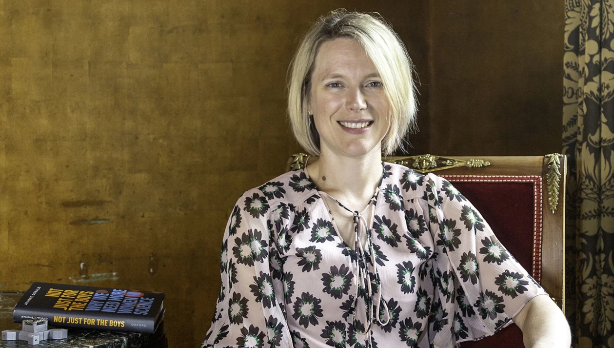
尽管卡文迪许实验室主攻物理学,目前,研究人员也在致力于揭开癌症的谜团。这其中包括同时在卡文迪许实验室和英国癌症研究中心(CRUK)剑桥研究所工作的莎拉·伯恩蒂克(Sarah Bohndiek)教授。
Although primarily associated with physics, today, researchers at the Cavendish are also helping to unravel the mysteries of cancer. This includes Professor Sarah Bohndiek, who is jointly based at the Cavendish and the CRUK Cambridge Institute.

莎拉·伯恩蒂克(Sarah Bohndiek)
Bohndiek’s team specialises in the use of light to uncover hidden properties of disease, especially cancer. They are developing devices that go beyond the visible spectrum, using different wavelengths of light to enable earlier detection.
该团队的一个关键项目涉及光声成像技术,这是一种结合光和超声波的技术。普通的超声扫描仪向体内发出声波,测量的是回声。而光声成像系统则测量的是当分子吸收光并升温时产生的超声波。
One of their key projects involves photoacoustic imaging, a technology combining light and ultrasound. Normal ultrasound scanners send sound into the body and measure the echo. Photoacoustic imaging systems, however, measure the ultrasound generated when molecules absorb light and heat up.
光声图像显示了体内分子的变化情况,尤其适用于测量血氧水平。伯恩蒂克教授团队的助理研究员托马斯·埃尔斯(Thomas Else)表示:“这一点对癌症监测很有用。由于癌细胞生长迅速,其含氧水平往往低于健康组织。”
Photoacoustic images show how molecules in the body are changing and are particularly useful for measuring oxygen levels in the blood. “This is a useful feature for looking at cancer,” said Thomas Else, a research associate in Bohndiek’s team. “Cancers tend to have lower oxygen levels than healthy tissue because they grow so quickly.”
目前最常用于检测和监测癌症的设备,如核磁共振成像(MRI)和乳腺X射线检查机器,都笨重且昂贵,另外乳腺X光检查使用了电离辐射,患者需要承受挤压乳房带来的疼痛。
Most widely-used methods for detecting and monitoring cancer – such as MRI and X-ray mammography machines – are bulky and expensive pieces of equipment, and in the case of mammography uses ionising radiation and applies painful compression.
“光声成像不使用电离辐射,这使其更安全且适合长期监测,并且可以像超声波一样在床边生成高对比度图像,”埃尔斯说,“光声系统的成本也比MRI机器低,机器也更便携,这使其拥有更广阔的医疗应用场景,成为一种有价值的癌症监测技术。”
“The lack of ionising radiation makes photoacoustic imaging safe and suitable for long-term monitoring, and it can produce high-contrast images at the bedside, like an ultrasound,” said Else. “Photoacoustic systems are also lower cost and more portable than MRI machines, which makes it a valuable cancer monitoring technique in a wider range of medical settings.”
光声成像已经在欧洲获得CE认证,并在美国获得FDA批准用于乳腺癌检测,这可能成为替代传统乳腺X线的一个高性价比方案。
Photoacoustic imaging is already CE-marked in Europe and FDA-approved in the United States for breast cancer detection which could be a cost-effective alternative to traditional mammograms.
然而,与其他基于光的医疗技术一样,光声成像在应用中面临着一个挑战:它在肤色较深的人群中检测表现不佳。影响肤色的主要色素——黑色素会吸收光,这意味着能够通过皮肤被测量到的光会减少。
However, one of the challenges with the application of photoacoustic imaging, like several light-based medical technologies, is that it does not perform as well for people with darker skin tones. Melanin, the main pigment that affects skin tone, absorbs light, meaning less light can get through the skin to make a measurement.
通常,光声成像使用的是从可见光谱的红色端到红外线的光波,波长大约在700到900纳米之间。在这个范围内,血液中的血红蛋白是主要的对比源。但在肤色较深的患者中,黑色素可能会掩盖血红蛋白的信号。
Typically, photoacoustic imaging uses light from the red end of the visible spectrum to the infrared – at wavelengths around 700 to 900 nanometres. In this range, haemoglobin in the blood is the main source of contrast. But in darker-skinned patients, melanin can overwhelm the haemoglobin signal.
“皮肤中的黑色素会阻挡一些光线,阻碍其深入组织并到达我们想要显像的癌症或器官,”埃尔斯说,“我们希望研究基于光的成像技术在有色人种中表现不佳的物理原因,这可能有助于我们解决这一问题。”
“Melanin in the skin blocks some light and prevents it from getting deep into the tissue and to the cancer or the organ we want to image,” said Else. “We wanted to look at the physics behind the underperformance of light-based imaging technologies for people of colour, which could help us address it.”

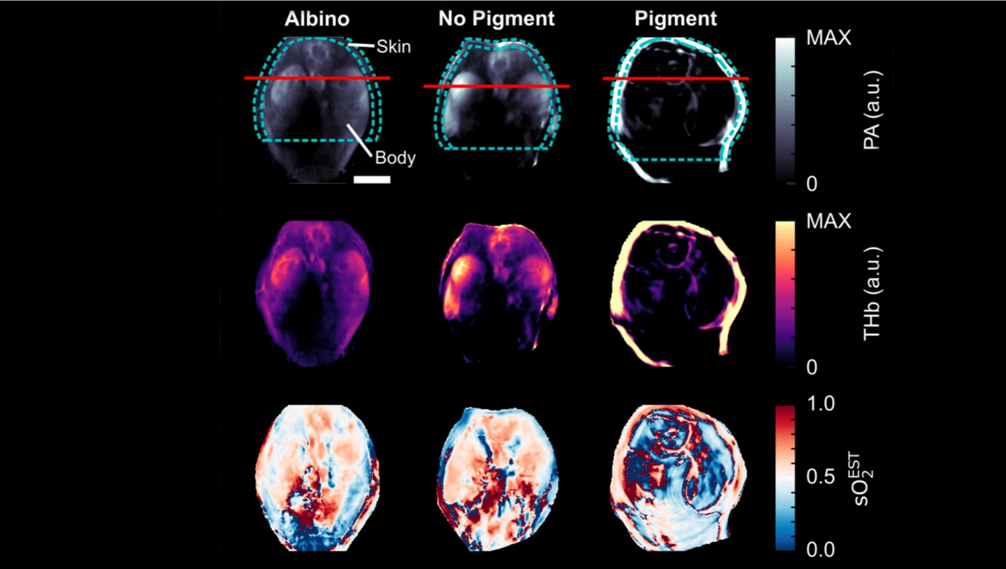
白化小鼠和有色小鼠脾脏和肾脏的光声图像
Photoacoustic images of mouse spleen and kidneys in albino and pigmented mice
在最近发表在《生物医学光学杂志》上的一项研究中,埃尔斯和他的同事们使用具有不同色素沉着程度的皮肤计算机模型来模拟光声图像。他们在具有不同色素沉着模式的小鼠和模拟不同肤色的凝胶模型(称为“幻影”)中验证了这些结果。
In a study recently published in the Journal of Biomedical Optics, Else and his colleagues used a computer model of skin with varying levels of pigmentation to simulate photoacoustic images. They validated the results in mice with different pigmentation patterns and in gel models, called phantoms, that mimic different skin tones.
“我们发现了两点主要影响因素:其一,如果你试图在肤色较深的人群中测量癌症检测的关键指征——血氧含量,你会得到不准确的测量结果,”埃尔斯说,“相较于白人患者,黑人患者血氧含量会被高估。你还会得到质量较低的图像,图像中会有噪声和变形,可能导致临床医生对扫描结果的解读发生偏差,肿瘤的攻击性很有可能看起来比实际更强。‘’
“We found two main effects: one was if you try to measure blood oxygenation – which is important in cancer – in people with dark skin, you get incorrect measurements,” said Else. “Blood oxygenation in Black patients is overestimated compared to white patients. You also get lower quality images, with noise and distortions in the image, that could lead a clinician to misinterpret the scan, potentially making a tumour look more aggressive than it really is.”
为了应对这些差异,研究人员正在与癌症早期检测联盟合作,与阿登布鲁克医院的志愿者一起进行进一步研究,目的在于完善这项技术并确保其在不同肤色中的有效性。
To address these discrepancies, the researchers are conducting further studies with volunteers at Addenbrooke’s Hospital in collaboration with the Alliance for Cancer Early Detection to refine the technology and ensure its efficacy across diverse skin tones.
伯恩蒂克教授的团队还在探索使用光声技术来定制放射治疗。通过使用光声技术来了解癌症对治疗的反应,由此临床医生可更有效地定制辐射剂量。
Bohndiek’s team is also exploring the use of photoacoustics for tailoring radiotherapy. By using photoacoustics to understand how cancers respond to treatment, clinicians might be able to customise radiation doses more effectively.
研究人员还在探索其他光基技术,如脉搏血氧仪和智能手表,这些设备在黑人患者中的血氧读数都存在偏差。他们的目标是开发能够不受肤色影响提供准确测量的可穿戴设备。
The researchers are also exploring other light-based technologies, like pulse oximeters and smartwatches, which have shown biases in blood oxygenation readings for Black patients. They aim to develop wearable devices that provide accurate measurements regardless of skin tone.
只要直面解决这些偏差,研究人员相信他们可以创造出更好、更具包容性的医疗设备,这不仅可以改进癌症的检测和监测,还能提升医疗技术的整体质量和可及性。
By tackling these biases head-on, the researchers believe they can create better and more inclusive medical devices that not only improve cancer detection and monitoring but also enhance the overall quality and accessibility of healthcare technology.
埃尔斯说,“虽然我们致力于开发和改进技术,但我们的关注点始终如一:我们希望改进癌症的检测和监测,让每位患者都有最大的机会。”
“Although we’re working on developing and improving technologies, our focus remains the same: we want to improve cancer detection and monitoring, so that every patient is given the best possible chance,” said Else.
莎拉·伯恩蒂克(Sarah Bohndiek)是剑桥大学基督圣体学院的教员。托马斯·埃尔斯(Thomas Else)隶属于剑桥大学克莱尔学院。
Sarah Bohndiek is a Fellow of Corpus Christi College, Cambridge. Thomas Else is a Member of Clare College, Cambridge.

 中文
中文 English
