Advanced MRI scans help identify one in three concussion patients with ‘hidden disease’
“脑震荡是影响成年人的第一大神经系统疾病,这就是为什么我们需要一种方法来识别那些最有可能出现持续症状的患者。”
——弗吉尼亚·纽康姆
“Concussion is the number one neurological condition to affect adults, which is why we need a way of identifying those patients at greatest risk of persistent symptoms”
——Virginia Newcombe
剑桥大学的研究人员表示,为脑震荡患者提供一种名为弥散张量成像MRI的脑部扫描,有助于识别出每三位患者中会出现可能影响生活的持续症状的那一位。
Offering patients with concussion a type of brain scan known as diffusion tensor imaging MRI could help identify the one in three people who will experience persistent symptoms that can be life changing, say Cambridge researchers.
在欧洲每年大约每200人中就有一人遭受脑震荡。在英国,每年有超过100万人因近期头部受伤到急诊室就诊。脑震荡已成为全球最常见的脑损伤形式。
Around one in 200 people in Europe every year will suffer concussion. In the UK, more than 1 million people attend Emergency Departments annually with a recent head injury. It is the most common form of brain injury worldwide.

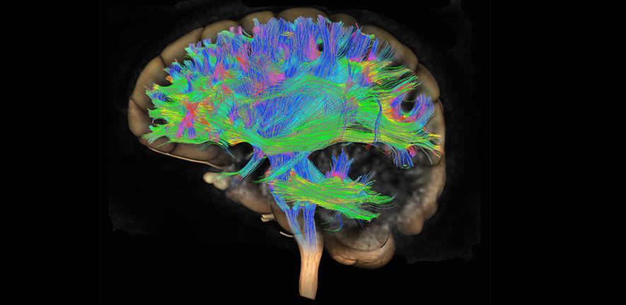
大脑弥散张量成像(DTI)
Diffusion tensor imaging (DTI) MRI of the human brain - stock photo
Credit: Callista Images (Getty Images)
当英国的患者因头部受伤前往急诊室时,医务人员会根据英国国家卫生和临床技术优化研究所(NICE)头部损伤指南进行评估。根据其症状,患者可能会被转诊进行CT扫描,以检查包括淤伤、出血和肿胀在内的脑损伤。
When a patient in the UK presents at an Emergency Department with head injury, they are assessed according to the NICE head injury guidelines. Depending on their symptoms, they may be referred for a CT scan, which looks for brain injuries including bruising, bleeding and swelling.
然而,CT扫描只能发现不到十分之一的脑震荡患者存在的异常,但在扫描后从急诊科出院的患者中有30-40%会出现持续数年且影响到生活的症状。这些症状包括严重疲劳、记忆力差、头痛和心理健康问题(包括焦虑、抑郁和创伤后应激障碍)。
However, CT scans identify abnormalities in fewer than one in 10 patients with concussion, yet 30-40% of patients discharged from the Emergency Department following a scan experience significant symptoms that can last for years and be potentially life-changing. These include severe fatigue, poor memory, headaches, and mental health issues (including anxiety, depression, and post-traumatic stress).
来自剑桥大学医学院和阿登布鲁克医院的重症监护及急诊科医生弗吉尼亚·纽康姆(Virginia Newcombe)博士表示:“大部分头部受伤的患者出院回家时会拿到一张单子,告知他们需注意的脑震荡后遗症,并告知他们如果症状加重,要寻求全科医生的帮助。”
Dr Virginia Newcombe from the Department of Medicine at the University of Cambridge and an Intensive Care Medicine and Emergency Physician at Addenbrooke’s Hospital, Cambridge, said: “The majority of head injury patients are sent home with a piece of paper telling them the symptoms of post-concussion to look out for and are told to seek help from their GP if their symptoms worsen.
“问题在于脑震荡的性质意味着患者和他们的全科医生,往往没有意识到他们的症状严重到需要随访。患者将其描述为一种‘隐性疾病’,不像骨折那样明显。由于没有脑损伤的客观证据(如扫描),这些患者在寻求帮助时往往感到他们的症状被忽视或忽略了。”
“The problem is that the nature of concussion means patients and their GPs often don’t recognise that their symptoms are serious enough to need follow-up. Patients describe it as a ‘hidden disease’, unlike, say, breaking a bone. Without objective evidence of a brain injury, such as a scan, these patients often feel that their symptoms are dismissed or ignored when they seek help.”
纽康姆博士及其同事发表在柳叶刀子刊《eClinicalMedicine》上的一项研究表明,一种称为弥散张量成像(DTI)的先进MRI形式,可以显著改善已接受常规CT脑扫描的脑震荡患者的现有预后模型。
In a study published in eClinicalMedicine, Dr Newcombe and colleagues show that an advanced form of MRI known as diffusion tensor imaging (DTI) can substantially improve existing prognostic models for patients with concussion who have been given a normal CT brain.
DTI测量水分子在组织中的移动方式,提供连接大脑不同部位的通路(称为白质纤维束)的详细图像。标准MRI扫描仪可以用于测量这些数据,这些数据可以用来根据不同大脑异常区域的数量计算DTI“得分”。
DTI measures how water molecules move in tissue, providing detailed images of the pathways, known as white matter tracts, that connect different parts of the brain. Standard MRI scanners can be adapted to measure this data, which can be used to calculate a DTI ‘score’ based on the number of different brain regions with abnormalities.
2014年12月至2017年12月期间,纽康姆博士及其同事研究了欧洲神经创伤疗效-脑损伤联合研究中心(CENTER-TBI)招募的1000多名患者的数据。患者中有38%未完全康复,这意味着出院后三个月他们的症状仍在持续。
Dr Newcombe and colleagues studied data from more than 1,000 patients recruited to the Collaborative European NeuroTrauma Effectiveness Research in Traumatic Brain Injury (CENTER-TBI) study between December 2014 and December 2017. 38% of the patients had an incomplete recovery, meaning that three months after discharge their symptoms were still persisting.
研究团队为153名接受DTI扫描的患者进行DTI评分。这显著提高了预后的准确性——目前的临床模型可在100例患者中正确预测69例预后较差,而DTI将这一比例提高到100例中的82例。
The team assigned DTI scores to the 153 patients who had received a DTI scan. This significantly improved the accuracy of the prognosis – whereas the current clinical model would correctly predict in 69 cases out of 100 that a patient would have a poorer outcome, DTI increased this to 82 cases out of 100.

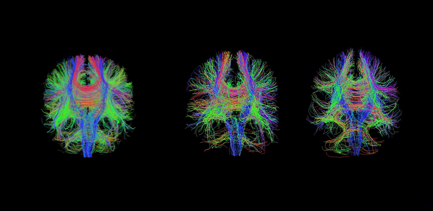
全脑弥散张量纤维束成像显示健康患者(左)和严重创伤性脑损伤后两天(中)和六周(右)的患者。
Whole brain diffusion tensor tractography showing healthy patient (left) and patient at two days (centre) and six weeks (right) after severe traumatic brain injury
研究人员还研究了血液生物标志物——头部受伤后释放到血液中的蛋白质——以了解这些蛋白质中是否有一种能提高预后的准确性。虽然仅靠生物标志物还不足够,但两个特定蛋白的浓度——受伤后12小时内的胶质纤维酸性蛋白( GFAP )和受伤后12至24小时内的神经丝轻链蛋白( NFL ) ——有助于识别出那些可能从DTI扫描中受益的患者。
The researchers also looked at blood biomarkers – proteins released into the blood as a result of head injury – to see whether any of these could improve the accuracy of the prognosis. Although the biomarkers alone were not sufficient, concentrations of two particular proteins – glial fibrillary acidic protein (GFAP) within the first 12 hours and neurofilament light (NFL) between 12 and 24 hours following injury – were useful in identifying those patients who might benefit from a DTI scan.
纽康姆博士表示:“脑震荡是影响成年人的第一大神经系统疾病,但卫生服务部门没有资源让每位患者定期复诊,这就是为什么我们需要一种方法来识别那些最有可能出现持续症状的患者。”
Dr Newcombe said: “Concussion is the number one neurological condition to affect adults, but health services don’t have the resources to routinely bring back every patient for a follow-up, which is why we need a way of identifying those patients at greatest risk of persistent symptoms.
“目前评估个体在头部受伤后预后的方法还不够好,但使用DTI——理论上任何配备MRI扫描仪的中心都可以实现——可以帮助我们做出更准确的评估。鉴于脑震荡症状可能对个人生活产生重大影响,这一点迫在眉睫。”
“Current methods for assessing an individual’s outlook following head injury are not good enough, but using DTI – which, in theory, should be possible for any centre with an MRI scanner – can help us make much more accurate assessments. Given that symptoms of concussion can have a significant impact on an individual’s life, this is urgently needed.”
研究团队计划对血液生物标志物进行更详细的研究,看看能否找到新方法来提供更简单、更实用的预测指标。他们还将探索将DTI应用于临床的方法。
The team plan to look in greater details at blood biomarkers, to see if they can identify new ways to provide even simpler, more practical predictors. They will also be exploring ways to bring DTI into clinical practice.
剑桥大学急诊医学NIHR临床讲师兼文章第一作者,、剑桥大学第一作者苏菲·里希特(Sophie Richter)博士补充道:“我们想看看当患者因脑损伤到医院就诊时,是否有方法可以整合获得的不同类型的信息——例如症状评估、血液检查和脑扫描——以改善我们对患者损伤及预后的评估。”
Dr Sophie Richter, a NIHR Clinical Lecturer in Emergency Medicine and first author, Cambridge, added: “We want to see if there is a way to integrate thedifferent types of information obtained when a patientpresents at hospital with brain injury – symptoms assessment, blood tests and brain scans, for example – to improve our assessment of a patient’s injury and prognosis.”
该研究由欧盟第七框架计划、惠康基金会和英国国家卫生与临床优化研究所资助。
The research was funded by European Union's Seventh Framework Programme, Wellcome and the National Institute for Health and Care Excellence.

 中文
中文 English
